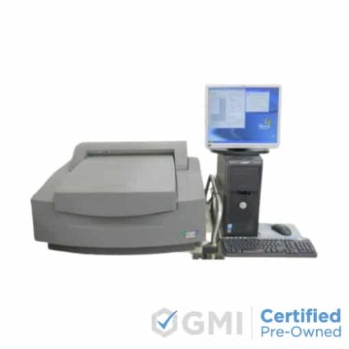
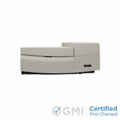
Product Description Versatile system handles gel sandwiches agarose and polyacrylamide gels membranes microplates and even microarrays. Powerful excitation sources and innovative high-quality confocal optics allow for the sensitive detection of low-abundance targets. Red- green- and blue-excitation wavelengths and a wide choice of emission filters enable imaging of an extensive variety of fluorophores. Automated four-color fluorescence scanning allows multiplexing of multiple targets in the same sample ensuring accuracy of analysis increasing throughput and saving time. Storage phosphor technology delivers high-resolution imaging and accurate quantitation of 3H 14C 125I 32P 33P 35S and other sources of ionizing radiation. Highly sensitive optics enable direct chemiluminescent imaging without intermediate exposure to films or screens. MOLECULAR DYNAMICS (GE Healthcare) Typhoon 9410 high performance gel and blot imager that can also perform image micro-arrays. Molecular Dynamics (GE) Typhoon 9410 Variable Mode Imager unites proven storage phosphor auto-radiography technology with four-color non-radioactive fluorescent labeling techniques. For DNA RNA and protein samples with the Molecular Dynamics Typhoon 9410you may choose from: storage phosphor auto-radiography direct blue-excited fluorescence (457 488 nm) direct green-excited fluorescence (532 nm) direct red-excited fluorescence (633 nm) chemiluminescence When one of the scanning modes is selected the Typhoon 9410 activates the appropriate optical components are automatically. Typhoon 9410 scans gels blots and mounted or unmounted storage phosphor screens up to 35 43 cm. The Typhoon 9410exhibits outstanding linearity quantitative accuracy and extremely low limits of detection. Choose from four different Typhoon models depending on your imaging needs. For added flexibility Typhoon 9200 9210 and 9400 can be upgraded as imaging needs change. All Typhoon models come with ImageQuant TL. Microarray Microarray Slide A micro-array slide spotted with cDNA from Micro-array ScoreCard control plate and cDNA from Incyte Genomics Inc. was hybridized with 25 pmol of Cy3-labeled skeletal muscle mRNA and Cy5-labeled liver mRNA. The slide was scanned on Typhoon 9410 at 10 &mu,m pixel size resolution. 2D Protien Staining Two Dimensional 2D- Protein Staining Two-dimensional 2-D protein gel stained with Deep Purple Total Protein Stain. Protein sample consists of HBL100 breast cell line and BT474 breast cell carcinoma. First dimension: Immobiline DryStrip pH 310 NL 24cm run on Ettan IPGphor IEF System. Second dimension: DALT Gel 12.5 run on Ettan DALTtwelve Large Vertical Electrophoresis System. Scanning: Typhoon 9400 Variable Mode Imager: Excitation green laser (532 nm), emission 560LP filter PMT 530 V 100 &mu,m resolution. The gel image shows the pH 38 region where most proteins are present. 4 Color Four-color competitive gel shift assay Four-color competitive gel shift assay to determine the relative affinities of the Salmonella spp. bacteriophage P22 repressor Mnt (1). In the assay the repressor is allowed to bind four different oligonucloetides labeled with FAM HEX TAMRA and ROX respectively at the same time. The intensities for each fluorophore in a lane are related directly to the relative affinity of the repressor protein for the oligonucleotide use In Lane Sizing In-lane sizing of PCR products In-lane sizing of PCR products. GeneScan -500 ROX size standard (yellow Applied Biosystems) Cy3-labeled PCR products (green) Alfexpress Cy5 Sizer 50500 (red GE Healthcare) and fluorescein-labeled PCR products (blue) were resolved in a 12% polyacrylamide gel glass sandwich References 1. Tsz-Kwong M. and Stormo G. D. Nucleic Acids Res. 29 24712478 (2001)
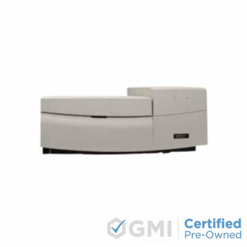
GE Amersham Molecular Dynamics Typhoon 9410 Молекулярный имидж-сканер
Доставка импортных компонентов по России от 3х недель транспортными компаниями СДЭК, Деловые Линии, Major Express.
Оплата физическими и юридическими лицами безналичным расчетом.
Цена может измениться после запроса у поставщика!